
Professor of Chemistry and Biochemistry
BIOGRAPHY
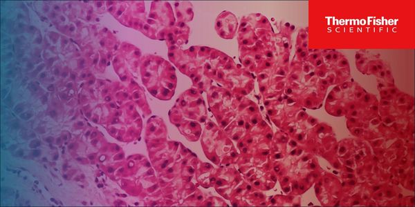
Deep Learning-Driven Image Analysis for Tracking Nucleolar Morphology
Presented at:
Immuno-Oncology Virtual Event Series 2026
Speakers
-

Leif Lindberg
Doctoral Candidate, DeRose Lab, University of OregonBIOGRAPHY -
Katelyn Alley, PhD
Researcher, DeRose Lab, University of OregonBIOGRAPHY


Abstract
Recent work has discovered that two platinum chemotherapeutics currently in widespread use operate by different cell death mechanisms. Cisplatin, particularly effective against testicular cancer, activates the DNA damage response. In contrast, oxaliplatin, a component of frontline colon cancer treatments, induces nucleolar stress and has also been linked to immunogenic cell death. The nucleolus is highly sensitive to chemotherapeutic stress by select agents, but its amorphous structure makes high-throughput analysis difficult and traditionally dependent on manual processing. We have developed expansion microscopy of the nucleolus to enable high-resolution structure analysis and monitor different states of reorganization upon drug treatments. For high-throughput screening analyses using conventional confocal microscopy, we leveraged Thermo Fisher Scientific’s Invitrogen™ SYTO™ RNASelect™ Red, a bright RNA selective dye that provides clear nucleolar staining. Using this dye, we developed deep learning models that accurately classify stressed versus unstressed cells and quantify nucleolar stress dependent morphological changes. This platform enabled us to track the rapid and irreversible onset of nucleolar stress induced by platinum chemotherapeutics, and to monitor the reversal of stress following treatment with the organic compound Actinomycin D.
Learning Objectives:
1. Understand how platinum chemotherapeutics effect nucleolar stress.
2. Use of an RNA specific dye to monitor nucleolar stress onset and reversal through expansion microscopy.
3. Understand deep learning models developed to describe the stress on cells when treated with platinum chemotherapies.
You May Also Like
APR 13, 2026 | 8:00 AM

Multiparametric flow-cytometry (MFC) is a diagnostic technique that allows a fluorescence-based assessment of surface, cytoplasmic and nuclear antigen expression on a suspension of blood, bo...
APR 14, 2026 | 11:00 AM

Join us for an insightful webinar exploring the transformative impact of ProTube™, Inpeco’s innovative solution designed to optimize the pre-pre-analytical phase in clinical labo...
APR 14, 2026 | 8:00 AM

Acute Myeloid Leukemia is the most common acute leukemia in adults affecting 2-6 adults per 100,000k globally. -With relapse rates as high as 50%, routine measurable residual disease (MRD)...
APR 16, 2026 | 6:00 AM
Physiologically relevant in-vitro liver systems are critical for translational research in metabolic diseases, including Metabolic Dysfunction-Associated Steatotic Liver (MASL) and Metabolic...
APR 16, 2026 | 8:00 AM

Early-stage product development strategies to meet regulatory requirements across different markets Differences in registration pathways across countries and corresponding strategies How to...
APR 20, 2026 | 12:00 AM
C.E. CREDITS

Webinar on coagulation QA basics covering PT/PTT lot rollover, geometric mean, heparin range, cumsum, AMR, troubleshooting, and accreditation standards....
Loading Comments...